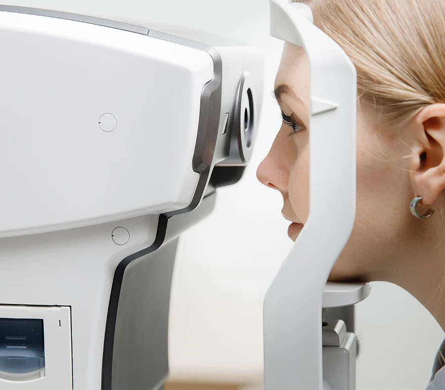
Optical Coherence Tomography
Optical coherence tomography, also known as OCT, is an imaging system that uses light waves to produce a high-resolution view of the cross-section of the retina and other structures in the interior of the eye.
Conditions Detected With an OCT
The images can help with the detection and treatment of serious eye conditions such as:
- Macular hole
- Macular swelling
- Optic nerve damage
- Age-related macular degeneration
- Macular pucker
- Glaucoma
- Cataracts
- Diabetic eye disease
- Vitreous hemorrhage
The OCT exam takes about 10 to 20 minutes to perform in your doctor’s office, and usually requires dilation of the pupils for the best results.
OCT uses technology that is similar to that of a CT scan of internal organs. With the scattering of light it can rapidly scan the eye to create an accurate cross-section. Each layer of the retina can be evaluated and measured and compared to normal, healthy images of the retina.
Contact Us
Contact us to today to learn more about Optical Coherence Tomography.
The doctors at Cincinnati Eye Institute have either authored or reviewed the content on this site.